
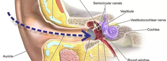
Deep in the external auditory canal, there is an eardrum as thin as a piece of paper, which can be said to be as thin as a cicada’s wing. The eardrum is the boundary between the outer ear and the middle ear. It isolates the middle ear from the outside world. Therefore, the middle ear and the external auditory canal are not connected in a normal ear. The eardrum is as thin as a cicada’s wing and translucent. Some people have a relatively straight external auditory canal. They can see the white eardrum with the help of light. In most cases, they need the help of special otological instruments to see it clearly.
The main function of the eardrum is to move inside and outside with the sound wave entering the external auditory canal, similar to the envelope of the drum. Because the eardrum is very thin, too much impact air flow, foreign objects and infection will cause eardrum rupture, which is often called tympanic membrane rupture or perforation. Like slapping. After eardrum rupture, if there is no infection and suppuration, it will generally heal by itself. The healing time varies from days to months. During this period, it is necessary to prevent water from entering the external auditory canal, avoid catching a cold, and avoid pinching the nose and puffing air (why will be discussed later). In addition to acute rupture, the eardrum will also appear local invagination due to various reasons. Most of them are the upper 1 / 3 part of the eardrum, which looks relatively loose, also known as the relaxation part, and the lower 2 / 3 part is the tension part. This part is like the envelope of the drum, which mainly plays the role of sound transmission. Perforation in this part will cause hearing loss. The invagination of the relaxation part will cause the exfoliated epithelial tissue to be unable to be discharged, so as to accumulate and grow continuously, which will compress inward and destroy the surrounding bones and structures, and also cause infection. This state is the formation of so-called cholesteatoma, which needs to be surgically removed as soon as possible.
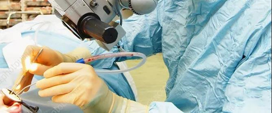
The perforation of tympanic membrane tension for more than three months is unlikely to be repaired and restored to normal state and function by relying on the healing ability of the human body. At this time, an otologist is needed to help with the so-called tympanic membrane repair operation. Although it is called tympanic membrane repair surgery, it does not repair the tympanic membrane as the name says. In fact, the doctor’s job is to enter the external auditory canal through an operating microscope or Otoendoscope, see the structure of the tympanic membrane, remove the scar tissue that has lost its growth ability at the edge of the perforation with a micro instrument, expose the normal three-layer structure of the tympanic membrane, and then use artificial materials or membrane like tissues elsewhere in the body, such as the fascia wrapping the temporal muscle, The fascia or adipose tissue or cartilage sheet wrapping the Tragus Cartilage builds a bridge at the perforation site, so that the skin and mucosal epithelial tissue in the three-layer structure of the previously freshly exposed tympanic membrane will crawl along the bridge, and finally close the tympanic membrane perforation. The bridging tissue will be destroyed and absorbed again in the later reconstruction process to become the tympanic membrane structure or directly participate in the tympanic membrane reconstruction. The success of tympanic membrane repair is related to the repair ability of the body, the ventilation function of the middle ear and postoperative nursing.

Beyond the eardrum, you enter the mysterious middle ear cavity, where the structure is complex, similar to the interior of a clock. But the eardrum is not an isolated layer of thin structure. In order to complete the transmission of sound, in addition to the bone wall (drum ring) fixed around the external auditory canal and the bone edge of the upper wall of the external auditory canal closing the bottom of the external auditory canal, the upper part of the eardrum is also wrapped with a hammer bone with a shape similar to that of a hammer. The handle of the hammer is wrapped by the eardrum, the head of the hammer is upward, and the inside is connected with the incus through ligament joints, The other end of the incus is connected with the stirrup shaped like a stirrup through joint ligaments.
The oval bone plate just seals the entrance of the inner ear through a circle of ligaments. The transmission system composed of the whole three auditory ossicles, which is called the ossicular chain in medicine, is located in the bony fossa in the middle and upper part of the inner side of the tympanic membrane. Otologists call this attic like space the upper tympanic chamber, in which two muscles, many ligaments and fascia fix this precise sound transmission system like a rope. When the eardrum receives the sound and impacts the internal and external activities, it will drive the internal and external movement of the malleus handle. The malleus head transmits the movement to the anvil and the anvil to the stapes. Finally, it will cause the internal and external piston movement of the stapes base plate and transfer the kinetic energy of the eardrum into the inner ear.
Ossicular chain is one of the most delicate structural systems of the human body. Its normal operation allows us to perceive the change of sound frequency from 20Hz to 20000hz and the change of sound loudness of 1dB. Of course, the normal operation of the ossicular chain requires the middle ear cavity to be in a normal pressure state and the middle ear mucosa to be in a normal state. Once the middle ear pressure or middle ear mucosa changes, such as eustachian tube dysfunction, tympanic membrane perforation and middle ear inflammation, it will affect the activity of the ossicular chain and cause discomfort symptoms, including ear tightness, hearing loss, tinnitus, etc. In order to avoid these situations, the human body has set up many protective measures. For example, in order to prevent fragile eardrum perforation, the human body hides the eardrum in the deep external auditory canal, surrounded by hard bones. In order to prevent water from entering, the external auditory canal presents a special curved shape. In order to prevent insects and dust from entering, cerumen glands are set up. In order to prevent the sudden change of middle ear pressure, the eustachian tube opening and closing balance pressure is set (the eustachian tube is a tube connecting the middle tympanic chamber and nasopharynx cavity as the name suggests).
In order to reduce the damage to the inner ear caused by the sudden strengthening of external sound, two muscles were set, the tensor tympani muscle fixed at the malleus neck and the stapes muscle fixed at the stapes neck. In addition to stabilizing the ossicular chain, the two muscles also have important protective effects. When the stapes muscle is stimulated by strong sound (such as thunder and talking loudly to the ear), it can contract, tighten the stapes base plate outward, reduce the activity of the stapes, and reduce the sound energy entering the inner ear. The tensor tympani muscle contracts as needed when the human body performs chewing and other actions, pulls the malleus handle to the inside, and the tympanic membrane is tense, which reduces the transmission of low-frequency sound generated by chewing action and improves the transmission of high-frequency sound, so that we can still effectively recognize the sound during chewing.